Who We Are
AOPLab, the Applied Optics and Polarimetry Lab, is a research team at LPICM, CNRS, École polytechnique and Institut Polytechnique de Paris, Palaiseau, France. We bring together expertise in various fields to build new polarimetric measurement systems that can be used in the laboratory as well as in clinical and field environments.
Our studies cover complete and partial imaging Mueller matrix polarimetry, both wide-field and microscopy, spectroscopic ellipsometry and spectroscopic Mueller polarimetry. We work on calibration techniques and surgical imaging instrumentation, simulate various scenarios of polarized light–tissue interaction, and implement image processing and AI-assisted segmentation. We specialize in designing reliable instruments where the hardware, calibration, acquisition strategies and data treatment algorithms are developed together in a coherent way.
Our future research is directed towards the creation of compact and fast intelligent polarimetric systems, including device miniaturization, real-time data processing, development of robust segmentation techniques, and instrument prototypes deployable in clinics and field environments. We aim to make polarimetric imaging easier to use, easier to interpret, and ready for meaningful translation.
A Journey Through Innovation
Tracing the development of our research group (recent milestones first) and its scientific foundations.
Next-Generation Wide-Field Imaging Polarimeter
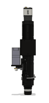
The next-generation wide-field imaging polarimetric system, designed and built at LPICM, combines a new miniaturized hardware design with an AI-based data processing and segmentation pipeline, ensuring near-video-rate streaming of polarimetric images. The system is designed to enable the translation of polarimetric imaging beyond laboratory settings.
PRISMM Hi! PARIS Funding
Hi! PARIS funds our PRISMM project "Polarimetric Real-time Imaging and Simulation using Mueller Matrix Imaging Platform for Neurosurgery" (2026–2028). This work supports the development of an AI-assisted real-time neurosurgical polarimetry workflow.

Plant Polarimetry
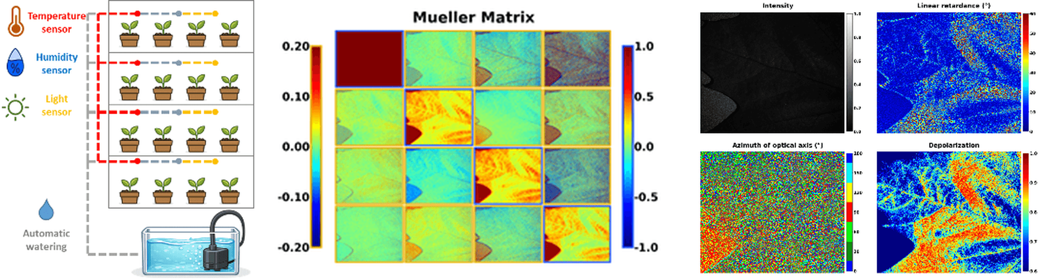
The application of wide-field imaging Mueller polarimetry for the assessment of plants' biotic and abiotic stress is becoming a new direction of our research. The measured polarimetric observables, combined with AI-based data post-processing, help read the microstructure of plant leaves and associated stress-linked changes without destructive sampling.
OptiSkin and MULTIFUSE ANR Funding
OptiSkin (2025–2029), "Multi-modality and multi-scale optical characterization and modelling of human skin applied to cancer diagnosis" (ANR-24-EXLU-0009), PEPR LUMA, France 2030. MULTIFUSE (2025–2028), "Advanced Multimodal Sensing and Data Fusion for Early Digital Detection of Plant Stress Symptoms", EIG CONCERT-Japan programme.


First In-Vivo Polarimetric Measurements during Neurosurgery
The team of the HORAO project performed the first feasibility study for end-to-end in-vivo brain tissue classification during brain tumor surgery using the wide-field imaging Mueller polarimeter, thus marking the transition from preclinical to clinical studies.
Institut Polytechnique de Paris Support
Institut Polytechnique de Paris supports the prospective studies on a real-time imaging Mueller polarimetric guidance system for neurosurgery by funding the PhD thesis work of S. Chae (2024–2027).
Campus France Partenariat Hubert Curien (PHC) Support
The PHAETHON Franco-Latvian PHC-OSMOSE project (2024–2025) supported the study on using photon time-of-flight measurements in brain tissues for medical diagnosis.

Mathematical Framework for Partial Mueller Polarimetry
Development of the theoretical mathematical framework for extracting the complete set of diagnostic polarimetric maps from partial 3×4 Mueller matrices that can be measured with a polarization-sensitive camera operating at video rate. This enables real-time polarimetric acquisition without measuring the complete 4×4 Mueller matrix, opening the avenue for in vivo intraoperative imaging (T. Novikova, J. C. Ramella-Roman, Opt. Lett. 47(21), 5549–5552 (2022)).
HORAO SNSF Sinergia Grant
The Swiss National Science Foundation awarded the HORAO Sinergia grant (2022–2026) to the international consortium, supporting polarimetric visualization of healthy brain fiber tracts for tumor delineation during neurosurgery. This collaboration brought together optical instrumentation, neurosurgery, neuropathology, and machine learning partners.

VECTOR Project École Polytechnique Funding
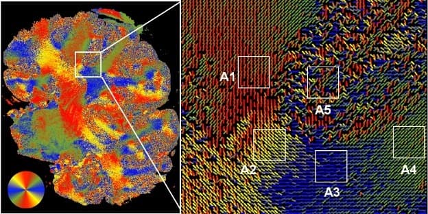
École polytechnique supported the VECTOR project (2020–2022), focused on proof-of-concept studies of the visualization of fiber bundles of healthy brain white matter and the delineation of brain tumor borders with wide-field imaging Mueller polarimetry during neurosurgery.
Campus France Partenariat Hubert Curien (PHC) Support
POLANNs, a Franco-New Zealand PHC Dumont d'Urville project (2019–2020), supported the development of neural-network-aided polarimetric diagnostics of cervical intraepithelial neoplasia.
MUSCAFE Project École Polytechnique Funding
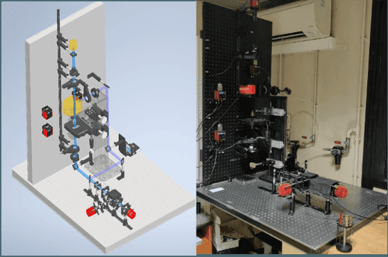
The MUSCAFE project (2019–2020) focused on building a Mueller microscope operating in both elastic scattering and fluorescence modes, in either reflection or transmission geometry, using commuting UV-A and visible light sources for the study of biological tissues.
BiCPIC Project — Chaire d'Alembert, Université Paris-Saclay Funding
The BiCPIC project "Binary Classification of Polarimetric Images for Cancer Diagnostics" (2017–2018) explored the implementation of the J-optimal Channelized Quadratic Observer for evaluating the detection performance of wide-field imaging Mueller polarimetry for cervical pre-cancer in tissue specimens. This study compared subsets of Mueller matrix data against the complete measurement, providing the mathematical groundwork for today's faster, partial Mueller polarimetry (M. Kupinski et al., Biomed. Opt. Express 9(11), 5691–5702 (2018)).
Physical Interpretation of Polarimetric Image Contrasts
Physical interpretation of experimental Mueller matrix image contrasts between healthy and cancerous human tissues paved the way for tissue optical biopsy. This breakthrough connected polarimetric observables directly to tissue microstructure and pathological status (T. Novikova et al., Appl. Phys. Lett. 102, 241103 (2013)).
MuellerFourier ANR Grant
The ANR-funded MuellerFourier project (2009–2011) explored the potential of using a custom-built back-focal-plane Mueller microscope for overlay measurements in microelectronics, strengthening the lab's position as a leader in Mueller polarimetric instrumentation and optical metrology.
Development of Wide-Field Imaging Polarimetry
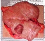
Moving from spectroscopic liquid-crystal-based Mueller polarimetry to multi-spectral liquid-crystal-based wide-field imaging Mueller polarimetry enabled the creation of spatial maps of the polarimetric properties of a sample. This opened the possibility of diagnostic characterization of complex tissue samples for various biomedical applications.
Monte Carlo Model for Polarized Light Propagation through Anisotropic Scattering Media
Generalization of the polarized Monte Carlo simulation technique to model the propagation of polarized light through anisotropic scattering biological tissues enabled the quantitative modelling of Mueller matrix images and derived polarimetric parameters of biological tissue. This provided the theoretical foundation for interpreting polarimetric contrast in structured media such as the white matter of the brain.
Mueller Polarimetry for Metrology in Microelectronics
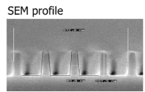
The development of a spectroscopic liquid-crystal-based Mueller polarimeter, combined with an in-house algorithm for solving the inverse problem of Mueller polarimetry, gave rise to metrological applications in microelectronics, including the estimation of critical dimensions and overlay metrology, opening an avenue for fast, non-destructive characterization of metrological structures with better accuracy than the standard scatterometry approach. The instrument design was commercialized by Horiba Jobin Yvon; several hundred instruments were sold worldwide.
Eigenvalue Calibration Method
LPICM members E. Compain, S. Poirier, and B. Drévillon published the seminal paper "General and self-consistent method for the calibration of polarization modulators, polarimeters, and Mueller-matrix ellipsometers" in Applied Optics 38(16), 3490–3502 (1999). Since then, the eigenvalue calibration method has become the gold standard for calibrating complete Mueller polarimetric instruments.
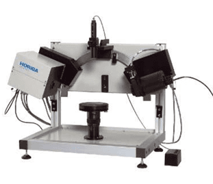
UVISEL Phase-Modulated Spectroscopic Ellipsometer
The phase-modulated spectroscopic ellipsometer UVISEL traces its origins to LPICM, where the underlying technology was developed and subsequently licensed to Jobin Yvon for commercialization. It is one of the LPICM-born polarimetric instruments that have gone on to become industry-standard tools in optical metrology worldwide.