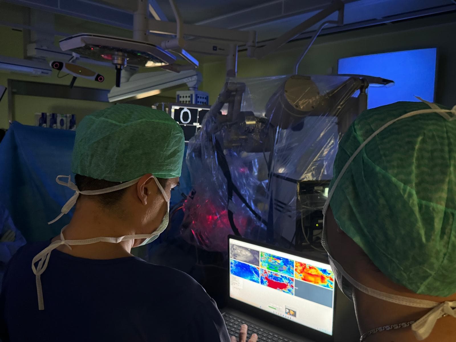
Surgery is the decisive step in brain cancer treatment, yet even with modern surgical microscopes the border between healthy and tumor tissue is very hard to see. The visual contrast between tumor and tumor-free tissue is low, but there is a clear structural difference between them. White matter of healthy brain, composed of densely packed fiber bundles of myelinated axons, represents an optically anisotropic medium. Tumor cells grow chaotically and destroy or displace fiber bundles, so optical anisotropy is lost within the tumor zone. AOPLab designs and builds wide-field imaging Mueller polarimetric systems that can visualize the optical anisotropy of biological tissue and make the boundary between fiber-preserved and fiberless zones visible in real time. This optical modality is label-free and does not disrupt the surgical workflow. The prototype developed within the framework of our prior HORAO project was successfully tested both ex vivo and in vivo. We are now developing the next generation of more accurate, compact, and faster polarimetric instruments for brain, spine, head, and neck surgery.
The Clinical Challenge
Brain tumor surgery pursues a delicate balance: remove as much tumor tissue as possible while protecting the white matter fiber bundles, which control movement, speech, and vision. Under white-light illumination the boundary between tumor and tumor-free tissue is hardly visible, so surgeons risk either leaving residual tumor cells (the main cause of recurrence) or damaging functional fiber tracts (the main cause of postoperative neurological deficit).
Seeing the Hidden Architecture of Brain White Matter
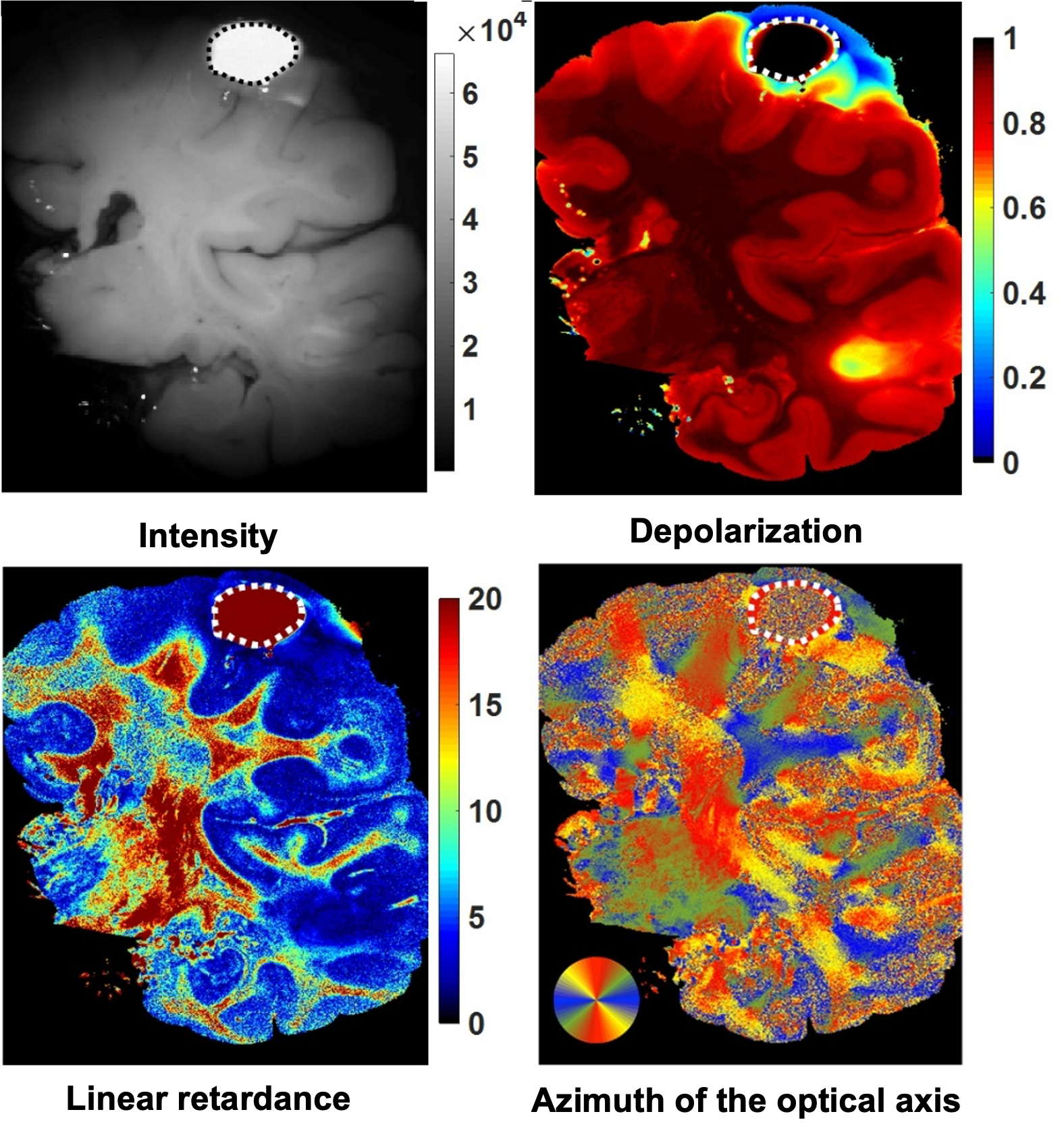
Healthy brain white matter is made of densely packed, aligned bundles of myelinated axons, causing so-called form birefringence. The optical axis of such a linearly birefringent medium is correlated with the fiber bundle direction. Our custom-built wide-field imaging Mueller polarimeter measures the full 4×4 Mueller matrix images and, by applying a pixel-wise non-linear data compression algorithm, namely Lu–Chipman decomposition (S.-Y. Lu and R. A. Chipman, J. Opt. Soc. Am. A 13(5), 1106–1113 (1996), 10.1364/JOSAA.13.001106), produces the maps of depolarization, linear retardance, and the azimuth of the optical axis of brain tissue.
Distinguishing Brain Tumor from Tumor-Free Brain Tissue
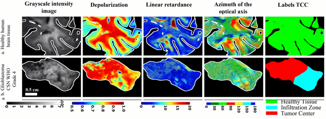
Brain tumor growth destroys the ordered architecture of healthy brain white matter, and tumor tissue loses optical anisotropy. All polarimetric properties of the brain are affected: depolarization and linear retardance values drop, and the azimuth of the optical axis takes random values within the tumor zone. Across healthy tissue and glioblastoma (WHO grade 4) specimens, the values of depolarization and retardance track the transition from healthy parenchyma, through the infiltration zone, to the tumor core, matching the histological ground truth. This is the structural contrast missing in white-light images, and the signal our segmentation models learn to use.
First Polarimetric Measurements of Brain In Vivo
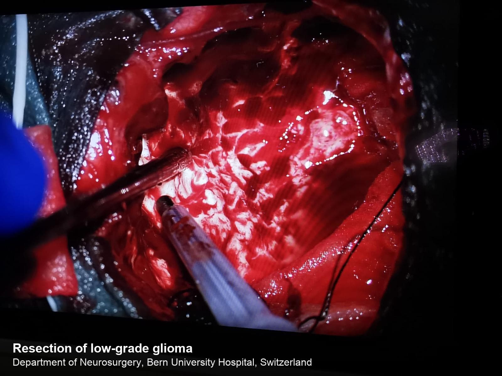
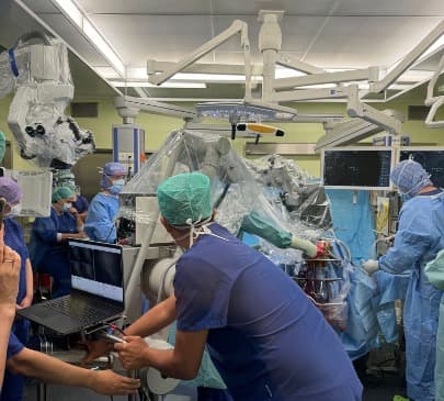
After polarimetric imaging pipeline validation on hundreds of ex vivo specimens, a new miniaturized Mueller polarimetric system was deployed during live brain tumor resection, producing the first in vivo polarimetric measurements of that kind. This marks the beginning of the transition of wide-field imaging Mueller polarimetry from the optical bench to the operating room, and a key step toward label-free, real-time visualization of brain tumor and fiber tracts in tumor-free brain tissue during neurosurgery.
Related Publications
Gros, É., Sakiri, A., Moriconi, S., Jankauskas, A., Hasler, D., Lucas, T., McKinley, R., Maragkou, T., Novikova, T., Schucht, P., Rodríguez-Núñez, O.
Brain Tissue Polarimetry Under Simulated Intraoperative Conditions: Effects of Edema and Temperature
Journal of Innovative Optical Health Sciences·2026
Chae, S., Wang, M., Lucas, T., Huang, T., Albano, G., Rodríguez-Núñez, O., Pierangelo, A., Gros, É., Maragkou, T., McKinley, R., Schucht, P., Novikova, T.
Biomedical Optics Express·2026
Tuchin, V. V., Novikova, T., Wang, L. V., Zimnyakov, D. A., Ma, H., Alonova, M. V., Wan, J.
Optical Polarization in Biomedical Applications (2nd Edition)
Springer Series in Biophysics, Springer Berlin Heidelberg New York·2026
Novikova, T., Chae, S., Lucas, T., Rodríguez-Núñez, O.
Polarized-light microscopy
Optical Techniques in Clinical Applications, Springer Nature·2026
Wang, M., Chae, S., Lucas, T., Rodríguez-Núñez, O., Gros, É., Hahne, C., Maragkou, T., McKinley, R., Schucht, P., Novikova, T.
Proc. SPIE 13854, 1385405·2026
Diamant, A., Lucas, T., Rodríguez-Núñez, O., Chae, S., Gros, É., Hahne, C., Maragkou, T., McKinley, R., Schucht, P., Novikova, T.
Proc. SPIE 13854, 138540F·2026
Oberti, N., Rodríguez-Núñez, O., Hasler, D., Schucht, P., McKinley, R., Novikova, T., Maragkou, T., Gros, É.
Proc. SPIE 14098, Tissue Optics and Photonics IV, 140981H (SPIE Photonics Europe)·2026
Lucas, T., Rodríguez-Núñez, O., Iqbal, A., Chae, S., Gros, É., Hahne, C., Maragkou, T., McKinley, R., Schucht, P., Novikova, T.
Proc. SPIE PC14094, Biomedical Spectroscopy, Microscopy, and Imaging IV, PC1409405 (SPIE Photonics Europe)·2026
Rodríguez-Núñez, O., Gros, É., Lucas, T., Chae, S., Hahne, C., Hasler, D., Maragkou, T., McKinley, R., Novikova, T., Schucht, P.
Proc. SPIE PC13841, Multimodal Biomedical Imaging XXI, PC1384107 (SPIE Photonics West)·2026
Rodríguez-Núñez, O., Lucas, T., Gros, É., Baier, D., Chae, S., Hahne, C., Hasler, D., Maragkou, T., McKinley, R., Schucht, P., Novikova, T.
Proc. SPIE 13854, Polarized Light and Optical Angular Momentum for Biomedical Diagnostics 2026 (SPIE Photonics West)·2026
Wang, M., Chae, S., Lukinsone, V., Lucas, T., Rodríguez-Núñez, O., Gros, É., Hahne, C., Maragkou, T., McKinley, R., Schucht, P., Novikova, T.
Biomedical Optics Express, 16(12), 5261–5278·2025
Hahne, C., Rodríguez-Núñez, O., Gros, É., Lucas, T., Novikova, T., Maragkou, T., Schucht, P., McKinley, R.
Physically Consistent Image Augmentation for Deep Learning in Mueller Matrix Polarimetry
IEEE Transactions on Image Processing, 34, 6953–6962·2025
Hahne, C., Diaz, I., Rodríguez-Núñez, O., Gros, É., Lucas, T., Novikova, T., Maragkou, T., Schucht, P., McKinley, R.
Polarimetric Feature Analysis of Mueller Matrices for Brain Tumor Image Segmentation
Optics Express, 33(20), 43379·2025
Gros, É., Rodríguez-Núñez, O., Moriconi, S., McKinley, R., Hewer, E., Lucas, T., Vassella, E., Schucht, P., Novikova, T., Hahne, C., Maragkou, T.
Alignment of Histological and Polarimetric Large-Scale Imaging for Brain Tissue Characterization
Journal of Biomedical Optics, 30(9), 096003·2025
Novikova, T.
Imaging Mueller polarimetry for biomedical diagnosis: Recent developments
EPJ Web of Conferences, 335, 04007·2025
Novikova, T.
Imaging Mueller Polarimetry: a New Lens on Human Health
Optica Biophotonics Congress 2025, Technical Digest Series, paper TTu3F.1·2025
Maragkou, T., Gros, R., Rodríguez-Núñez, O., Felger, L., Moriconi, S., McKinley, R., Novikova, T., Schucht, P., Pierangelo, A., Vassella, E., Hewer, E.
Diagnosis of Brain Tumors Using Wide-Field Imaging Mueller Polarimetry
Journal of Neuropathology & Experimental Neurology, 83(6), 409·2024
Gros, R., Rodríguez-Núñez, O., Felger, L., Moriconi, S., McKinley, R., Pierangelo, A., Novikova, T., Vassella, E., Schucht, P., Hewer, E., Maragkou, T.
IEEE Transactions on Medical Imaging, 43(12), 4120·2024
Moriconi, S., Rodríguez-Núñez, O., Gros, R., Felger, L. A., Maragkou, T., Hewer, E., Pierangelo, A., Novikova, T., Schucht, P., McKinley, R.
Near-real-time Mueller polarimetric image processing for neurosurgical intervention
International Journal of Computer Assisted Radiology and Surgery·2024
Gros, R., Rodríguez-Núñez, O., Felger, L., Moriconi, S., McKinley, R., Pierangelo, A., Novikova, T., Vassella, E., Schucht, P., Hewer, E., Maragkou, T.
Proc. SPIE 13010, Tissue Optics and Photonics III, 130100F·2024
Ivanov, D., Si, L., Felger, L., Maragkou, T., Schucht, P., Ma, H., Schanne-Klein, M.-C., Ossikovski, R., Novikova, T.
Journal of Biomedical Optics, 28(10), 102908·2023
Gros, R., Rodríguez-Núñez, O., Felger, L., Moriconi, S., McKinley, R., Pierangelo, A., Novikova, T., Vassella, E., Schucht, P., Hewer, E., Maragkou, T.
Effects of formalin fixation on polarimetric properties of brain tissue: fresh or fixed?
Neurophotonics, 10(2), 025009·2023
Felger, L., Rodríguez-Núñez, O., Gros, R., Maragkou, T., McKinley, R., Moriconi, S., Murek, M., Zubak, I., Novikova, T., Pierangelo, A., Schucht, P.
Biomedical Optics Express, 14(5), 2400–2415·2023
Yashin, K., Novikova, T., Shcheslavskiy, V.
Editorial: Optical imaging and Laser technologies in Neuro-Oncology
Frontiers in Oncology, 12, 1103711·2023
Moriconi, S., Felger, L., Rodríguez-Núñez, O., Gros, R., Hewer, E., Maragkou, T., Novikova, T., Pierangelo, A., Murek, M., Schucht, P., McKinley, R.
Towards Real-time Integration of Polarimetric Image-processing for Neurosurgical Applications
Brain and Spine, 3, 102159·2023
Rodríguez-Núñez, O., Gros, R., Felger, L., Maragkou, T., McKinley, R., Moriconi, S., Novikova, T., Pierangelo, A., Schucht, P.
Polarimetric markers for delineation of lesional margins during brain tumor surgery
Brain and Spine, 3, 102160·2023
Felger, L., Gros, R., Maragkou, T., McKinley, R., Moriconi, S., Murek, M., Novikova, T., Pierangelo, A., Rodríguez-Núñez, O., Zubak, I., Schucht, P.
Proc. SPIE 12364, 1236406·2023
Gros, R., Felger, L., Maragkou, T., McKinley, R., Moriconi, S., Murek, M., Novikova, T., Rodríguez-Núñez, O., Schucht, P., Zubak, I., Pierangelo, A.
Proc. SPIE PC12382, PC1238203·2023
McKinley, R., Felger, L., Gros, R., Hewer, E., Maragkou, T., Moriconi, S., Murek, M., Novikova, T., Rodríguez-Núñez, O., Pierangelo, A., Schucht, P.
Proc. SPIE PC12382, PC1238208·2023
Moriconi, S., Felger, L., Gros, R., Hewer, E., Maragkou, T., Murek, M., Novikova, T., Rodríguez-Núñez, O., Pierangelo, A., Schucht, P., McKinley, R.
Denoising diffusion networks applied to Mueller polarimetric images of brain tissue
Proc. SPIE PC12382, PC123820H·2023
Rodríguez-Núñez, O., Felger, L., Gros, R., Maragkou, T., McKinley, R., Moriconi, S., Murek, M., Pierangelo, A., Schucht, P., Zubak, I., Novikova, T.
Proc. SPIE PC12382, PC1238209·2023
Novikova, T., Rodríguez-Núñez, O.
Polarimetric techniques for the structural studies and diagnosis of brain
Advanced Optical Technologies, 11(5-6), 157–171·2022
Novikova, T., Pierangelo, A., Schucht, P., Meglinski, I., Rodríguez-Núñez, O., Lee, H. R.
Mueller Polarimetry of Brain Tissues
Polarized Light in Biomedical Imaging and Sensing, Springer·2022
McKinley, R., Felger, L., Hewer, E., Maragkou, T., Murek, M., Novikova, T., Rodríguez-Núñez, O., Pierangelo, A., Schucht, P.
Proc. SPIE 12136, 121360G·2022
Rodríguez-Núñez, O., Schucht, P., Hewer, E., Novikova, T., Pierangelo, A.
Brain fiber tracts imaging with polarized light: a potential navigation tool for tumor neurosurgery
Proc. SPIE PC11963, PC1196303·2022
Rodríguez-Núñez, O., Schucht, P., Hewer, E., Novikova, T., Pierangelo, A.
Polarimetric visualization of healthy brain fiber tracts under adverse conditions: ex vivo studies
Biomedical Optics Express, 12(10), 6674–6685·2021
Novikova, T., Schucht, P., Lee, H. R., Mezouar, M. H., Hewer, E., Raabe, A., Murek, M., Zubak, I., Goldberg, J., Kövari, E., Pierangelo, A.
Proc. SPIE 11641, Dynamics and Fluctuations in Biomedical Photonics XVIII, 1164105·2021
Rodríguez-Núñez, O., Schucht, P., Lee, H. R., Mezouar, M. H., Hewer, E., Raabe, A., Murek, M., Zubak, I., Goldberg, J., Kövari, E., Pierangelo, A., Novikova, T.
Proc. SPIE 11919, Translational Biophotonics: Diagnostics and Therapeutics, 119190Y·2021
Schucht, P., Lee, H. R., Mezouar, M. H., Hewer, E., Raabe, A., Murek, M., Zubak, I., Goldberg, J., Kövari, E., Pierangelo, A., Novikova, T.
IEEE Transactions on Medical Imaging, 39(12), 4376–4382·2020